-
An Excel file which lists all of the slides/images available in the Histology and Histopathology sets, grouped according to tissue type and pathology.

This work is licensed under a Creative Commons Attribution-NonCommercial-ShareAlike 4.0 International License.
Histology & histopathology
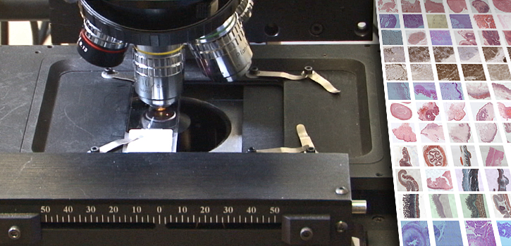
A collection of 320 annotated slides covering basic histology and histopathology.
Background and nature of the task
Presented through a virtual microscope, the histology and histopathology collection includes two sets (light-boxes) of normal tissues and ten sets of pathological sections grouped according to their tissue type. The entire collection constitutes 320 sections which are presented through a virtual microscope that models the functions of a conventional light microscope. Each section has an active descriptive legend that acts as a tutorial, by shifting the stage directly to the features described.
Instructions on how to use the microscope are given in the 'Help' file within the microscope. Background reading material on histology and microscopy is provided in the free OpenLearn course linked to below.
Histology, microscopy, anatomy and disease | OpenLearn - Open University
The slide sets were provided by a number of partners, including the Department of Pathology Cambridge, University College Hospital London, De Montfort University, the Institute of Ophthalmology, Milton Keynes NHS Trust and Leicester General Infirmary. Imaging and production of the virtual microscope was carried out at the Open University, as part of a digitisation project funded by JISC.
Duration and pattern of use
This activity may take 1-15 hours of work, depending on interest and experience.
You are not signed in to this website. More facilities might be available if you sign in.