Virtual petrographic microscope
Thin rock sections viewed under a polarising microscope.
Background and nature of the task
The many varieties of minerals in rocks can be identified by their optical properties in thin slices under a specialist polarising microscope – a branch of science known as petrography. This key skill of mineral identification is an essential part of rock classification, and a first step in understanding their origins. The microscope can also be used to investigate rock textures, microstructures and microfossils, giving insights into how the rock formed, and maybe deformed, and how it has changed through its history.
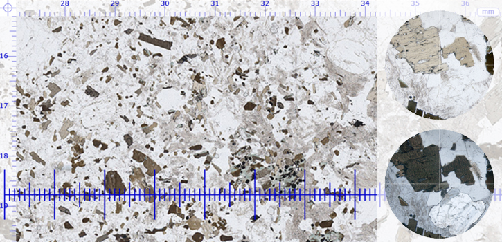
Polarized light interacts with slices of minerals just 30 microns (0.03 mm) thick to produce a dazzling array of optical effects – spectacular colours, delicate textures and intricate patterns. This beauty is more than skin deep, however: encrypted in these visual effects is a wealth of geological information. Learning how to uncover and use this information takes a great deal of practice, but this invaluable experience is readily gained with the Virtual Microscope.
Starting from a view of the whole thin section, you can zoom in either in steps or smoothly using a slider to view the smallest features (without breaking the glass slide!). Switch between plane-polarised and cross-polarised light to contrast their effects on the mineral crystals. Click on the rotating view hotspots to compare the same view side-by-side under plane and cross polars – then spin these round to see how the appearance of individual crystals changes. Add in some background information on the sample and some measurement tools and you have a powerful petrographic toolset to help you unlock the geological mysteries of any sample.
You are not signed in to this website. More facilities might be available if you sign in.