Using the digital microscope to count leukocytes in blood samples
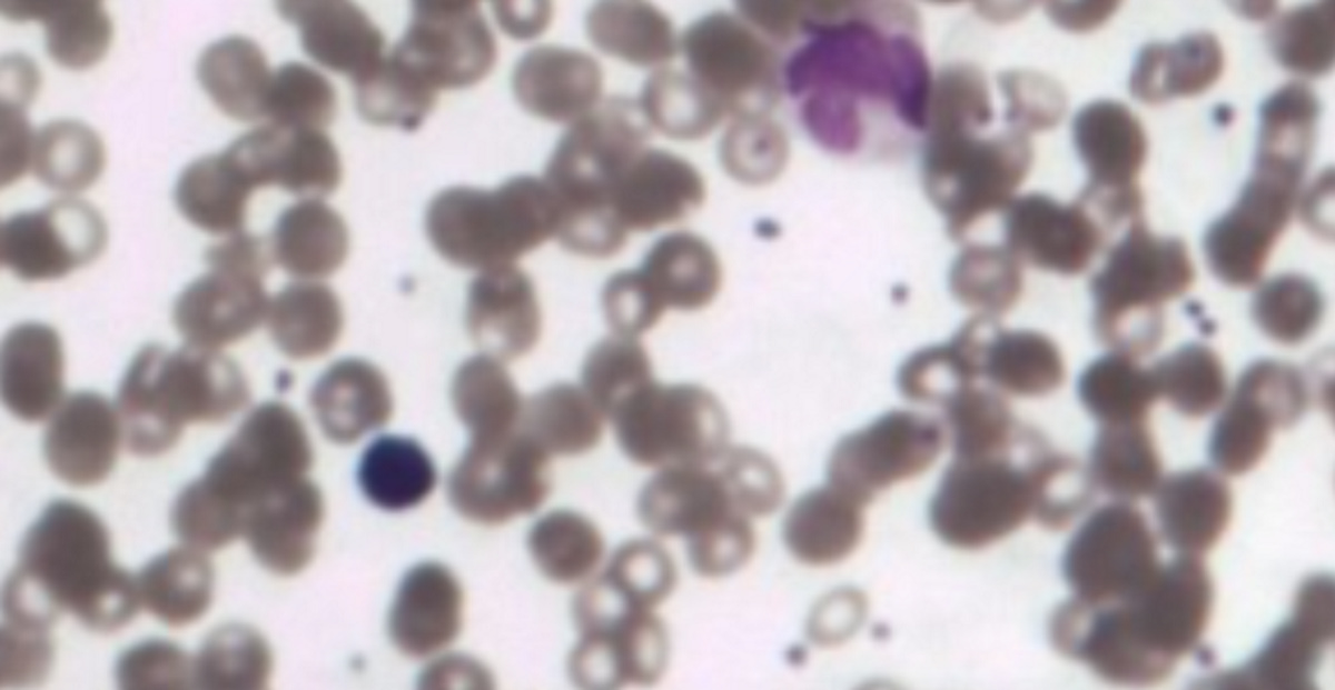
This activity allows you to use a digital microscope to test for
the presence of infection in blood samples. Two blood smear
samples are shown, one from a healthy person (termed the 'Normal' smear)
and a second from someone who is suspected to be fighting an infection (termed
the 'Leukocytosis' smear). The digital microscope can be used to compare the
number of leukocytes in each smear. The samples have been stained with a
blue/purple dye which specifically reveals the leukocytes in the
samples, as you can see in the image above where the leukocytes appear as a
distinct blue/purple colour.
You can choose a single track (labelled A-U) to count on each slide as a representative sample of the whole slide, or if you wish you can count all of the tracks on each slide. When you have obtained your counts for the normal and leukocytosis slides you should compare them. Is the count for the leucocytosis slide larger than the count for the normal slide? If so this suggests that the leukocytosis slide was taken from someone who was suffering from an infection.
Click on the link below to launch the digital microscope leukocyte counting activity.
You are not signed in to this website. More facilities might be available if you sign in.